Eating, Sleeping, Talking, Remembering: Which Part of the Brain Does What

Four things you almost certainly did today: ate something, slept, said something out loud, and remembered something. Each one is run from a different part of the brain, and the parts are easier to keep straight than the names suggest.
There is one trick that makes all of this stick, and it is worth knowing before the anatomy. Almost everything we know about which region does what came from watching what stopped working when that region was damaged. So you do not have to memorise a list of structures and jobs. You memorise what breaks, and the job follows from it.
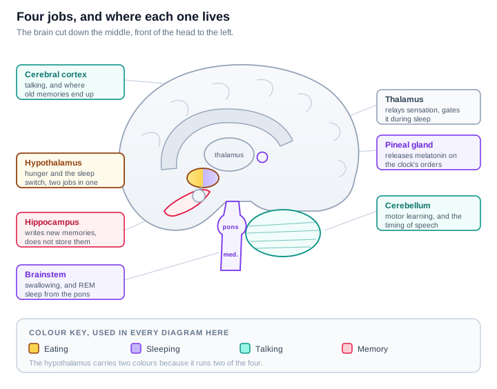
Before the detail, one orientation point. Two of these four are run from a lump of tissue weighing about four grams, tucked under the middle of the brain. The other two need the big folded outer layer. That split is most of the story.
Eating
The control centre is the hypothalamus, and within it a region called the arcuate nucleus does the sensing.
It reads two hormones that arrive in the blood and disagree with each other. Ghrelin comes from an empty stomach and means eat. Leptin comes from fat tissue and means there is fuel in store already. The arcuate nucleus holds two opposing populations of neurons, one that drives hunger and one that suppresses it, and your appetite at any moment is which population is currently winning.
Two neighbouring areas were the first clues, and they came from lesion experiments that are almost comically blunt.
| Area | Damage it | So its job was |
|---|---|---|
| Lateral hypothalamus | The animal stops eating | Saying go |
| Ventromedial hypothalamus | The animal overeats and becomes obese | Saying stop |
A rough way to keep them apart: damage the Lateral and you Lose weight, damage the Medial and you Mushroom.
Finishing a particular meal is handled lower down, in the brainstem. The vagus nerve reports stomach stretch to a region of the medulla called the nucleus of the solitary tract, which is why you feel full from volume and not only from calories. The medulla also runs swallowing itself, which is reflexive and not something the cortex is invited to supervise.
The cortex does get a say in wanting. Deciding that a particular food is worth having involves the reward circuitry and the orbitofrontal cortex, which is the difference between being hungry and specifically wanting chocolate.
Sleeping
The hypothalamus again, which is why it carries two colours in the map above.
Sleep needs two separate things: knowing what time it is, and having a switch.
The clock is the suprachiasmatic nucleus, roughly ten thousand neurons on each side, sitting directly above the point where the optic nerves cross. That position is not a coincidence. It receives a dedicated line from the retina, so it is set by light rather than by anything you decide. It then instructs the pineal gland to release melatonin when it gets dark. Melatonin is a signal that it is night, not a sedative.
The switch is the ventrolateral preoptic nucleus, which turns sleep on by actively inhibiting the arousal systems in the brainstem. Those arousal systems push back, which makes the two sides behave like a toggle rather than a dial, and is part of why you tend to be either asleep or awake rather than lingering in between.
Holding the switch steady is a third population, the orexin neurons of the lateral hypothalamus. The evidence here is unusually direct: in narcolepsy type 1 these neurons are largely gone. The original postmortem study found an 85 to 95 percent reduction, and later work has reported more. Without them the toggle flips when it should not, which is the condition.
There is a detail worth enjoying. Orexin was named for appetite, from the Greek for desire to eat, because that is what researchers first saw it do. Its more famous role turned out to be holding you awake. Hunger and wakefulness run through overlapping machinery, which is not surprising once you consider that an animal that sleeps through its chance to eat does not do well.
Once you are asleep, the pons takes over for REM sleep, generating it and also producing the muscle paralysis that stops you acting out dreams. Meanwhile the thalamus stops relaying sensory traffic to the cortex, which is why the room goes quiet even though your ears still work.
Talking
This is the one that is clearly lateralised. In roughly 95 percent of right-handed people, language lives in the left hemisphere, and the right hemisphere has little to say.
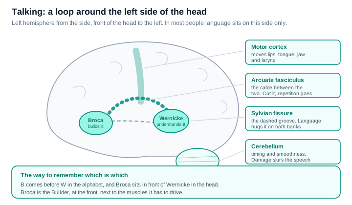
Two regions do the work, and the cleanest way to tell them apart is by what their damage sounds like.
| Damaged | How the speech sounds | Comprehension | Does the person notice? |
|---|---|---|---|
| Broca's area | Effortful, slow, stripped to key words | Largely intact | Yes, and it is frustrating |
| Wernicke's area | Fluent, well-paced, largely meaningless | Poor | Often not |
| The connection between them | Fluent and understood, but repetition fails | Intact | Yes |
That third row is the one that convinces people the model is real. If the two areas are intact but the arcuate fasciculus joining them is cut, someone can understand you perfectly and speak perfectly, yet cannot repeat back a phrase you just said. The parts work, the wire between them does not.
To remember which is which: B comes before W in the alphabet, and Broca sits in front of Wernicke in the head. Broca is the Builder, near the front, right next to the motor cortex it has to drive.
Two supporting players finish the job. The motor cortex actually moves the lips, tongue, jaw and larynx. The cerebellum handles timing and smoothness, which is why cerebellar damage produces slurred, poorly paced speech in someone whose language itself is intact.
Remembering
The headline structure is the hippocampus, curled into the temporal lobe on each side, and the single most important thing about it is counterintuitive.
The hippocampus does not store your memories. It writes them. Long-term storage ends up distributed across the cortex, and the hippocampus is the part that files new material into it. This is why hippocampal damage leaves old memories largely intact while making new ones impossible to form.
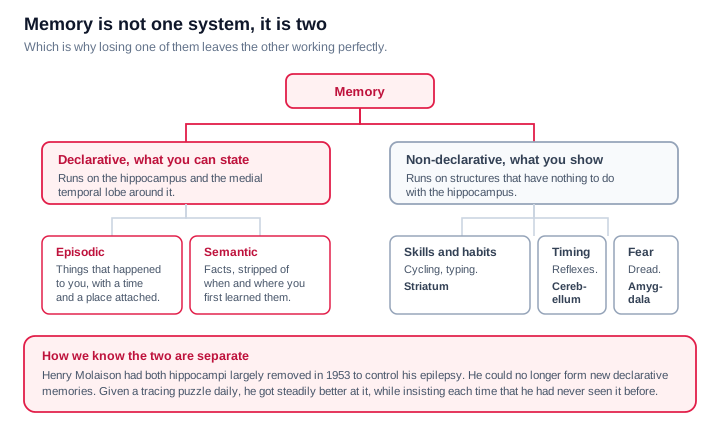
The case that established this is the most studied in neuroscience. In 1953 Henry Molaison had most of both hippocampi removed to control severe epilepsy. The seizures improved. He also lost the ability to form new conscious memories, for the remaining fifty five years of his life.
What makes his case decisive is not what he lost but what he kept. Given a tracing puzzle to practise, he got measurably better at it day after day, while insisting each time that he had never seen it before. His skill memory was recording perfectly. His memory of recording it was not. One kind of memory had been removed and the other was untouched, which told us they were never one system.
So memory divides into things you can state and things you can only show:
- Declarative, what you can state, runs through the hippocampus. It splits into episodic memory, events that happened to you with a time and place attached, and semantic memory, facts you know without remembering learning them.
- Non-declarative, what you show, runs elsewhere entirely. Skills and habits use the striatum. Conditioned timing and reflexes use the cerebellum. Fear learning uses the amygdala, which is why a frightening memory has a physical grip that a neutral one does not.
Holding a phone number in mind for the ten seconds it takes to dial it is a different thing again, called working memory, and it runs on the prefrontal cortex.
One connection back to the previous section: sleep is when recent material gets consolidated into longer-term cortical storage. Sleeping and remembering are not two unrelated items on this list.
The whole thing on one page
| Function | Main structure | Where it is | What its damage looks like |
|---|---|---|---|
| Hunger and fullness | Hypothalamus, arcuate nucleus | Deep middle, under the thalamus | Eating too little or too much |
| Ending a meal | Medulla, via the vagus nerve | Brainstem | Fullness from volume stops registering |
| Body clock | Suprachiasmatic nucleus | Hypothalamus, above the optic chiasm | Sleep timing unmoored from day and night |
| Falling asleep | Ventrolateral preoptic nucleus | Hypothalamus, front | The switch will not throw |
| Staying awake | Orexin neurons | Lateral hypothalamus | Narcolepsy |
| Dreaming sleep | Pons | Brainstem | REM lost, or acted out |
| Producing speech | Broca's area | Left inferior frontal | Effortful, telegraphic speech |
| Understanding speech | Wernicke's area | Left posterior temporal | Fluent speech that means little |
| Repeating speech | Arcuate fasciculus | Between the two | Repetition fails, everything else works |
| Clear articulation | Motor cortex and cerebellum | Precentral strip, and under the back | Slurred or mistimed speech |
| Forming new memories | Hippocampus | Curled into the temporal lobe | No new conscious memories |
| Skills and habits | Striatum | Deep, beside the thalamus | Skills stop improving with practice |
| Emotional weight of memory | Amygdala | In front of the hippocampus | Fear learning fails |
How to actually memorise this
Three hooks do most of the work.
Old brain, new brain. Eating and sleeping are housekeeping, and they are run from the hypothalamus and brainstem, deep and ancient and shared with every vertebrate. Talking and remembering need the cortex and the structures folded underneath it. If you can remember which of the four is housekeeping, you already know roughly where to look.
The hypothalamus is the housekeeper. Hunger, sleep, temperature, thirst, hormones. Four grams, running the parts of you that have to keep working whether or not you are paying attention.
Learn the breakages, not the boxes. Lateral hypothalamus, stops eating. Orexin neurons, narcolepsy. Broca, cannot build the sentence. Wernicke, cannot check it means anything. Hippocampus, cannot write it down. Every one of those is a story rather than a label, and stories are what stick.
Where this is simplified
Clean maps of the brain are always a little dishonest, and it is worth saying how.
These are not boxes, they are networks. No region works alone. Naming a structure for a function is shorthand for saying it is a necessary part of a network, not that the function lives inside it.
The Broca and Wernicke model is a useful simplification. It has been the standard teaching picture for over a century because the clinical patterns are so recognisable, but the modern understanding involves two processing streams and a wider set of regions, and the original areas do not map as neatly onto the symptoms as the classic story suggests. It is still the right first picture. It is not the final one.
People vary. Language is left-sided in most people but not all, and it is more often shared across both hemispheres in left-handers. Anatomy is a tendency, not a guarantee.
Damage rarely respects a boundary. Real strokes and injuries cut across several structures at once, so textbook-pure versions of these syndromes are less common than the textbooks make them look.
Sources and further reading
- Suprachiasmatic nucleus, neuron counts and function
- The suprachiasmatic nucleus, Current Biology review
- Reduced number of hypocretin neurons in human narcolepsy, Thannickal and colleagues
- Orexin deficiency in narcolepsy, 2025 review
- Hypocretin neuron loss in narcolepsy type 1, postmortem series
If you want the layer underneath this one, how the signals themselves travel, I wrote that up separately in The Human Nervous System: A Visual Guide.
Never miss a post
New guides on VMware Cloud Foundation, Aria Suite, and infrastructure automation. Follow the blog in your feed reader and new posts show up as soon as they are published.
Using a different reader? Copy the feed URL and add it there.