Cerebrospinal Fluid: Where It Comes From, Where It Goes, and What It Tells a Doctor

Your brain weighs about 1.4 kilograms and has roughly the consistency of set jelly. It is not built to hold its own shape against gravity, and nothing inside your skull is holding it up. It floats.
The fluid it floats in is cerebrospinal fluid, and it is doing considerably more than cushioning. You make around half a litre of it every day, you only ever hold about 150 mL at once, and the whole volume is replaced several times between waking up and going to bed.
Where it is made
Most of it comes from the choroid plexus, a frilly, highly vascular tissue that hangs inside the ventricles, the four fluid-filled chambers in the middle of the brain. Studies of isolated preparations put the choroid plexus at around 80 percent of total production, with the rest coming from the brain tissue itself.
This is not filtration. The choroid plexus actively pumps sodium across its epithelium, and water follows the salt. That distinction matters for two reasons. It means production continues more or less regardless of blood pressure, which is why a blockage keeps building rather than quietly equalising. And because it is an active transport process, a drug can slow it, which is how acetazolamide is sometimes used.
The epithelium also forms the blood-CSF barrier. The capillaries feeding the choroid plexus are leaky, but the cell layer wrapped around them is not, and it decides what gets through. The fluid that comes out is therefore not a blood filtrate. It is manufactured to specification.
| CSF | Blood plasma | |
|---|---|---|
| Protein | 15 to 45 mg/dL | around 7,000 mg/dL |
| Glucose | roughly two thirds of the blood level | reference |
| White cells | 0 to 5 per µL | thousands per µL |
| Red cells | none | billions per µL |
| Appearance | clear, like water | opaque |
That protein column is the one to look at. CSF carries a few hundred times less protein than plasma. It is an exceptionally clean fluid, and that cleanliness is what makes it such a good diagnostic sample, because almost anything you find in it that should not be there is a finding.
Where it goes
The route is a one-way circuit, not a loop. It is made deep in the middle, works its way down and out through a series of progressively narrower openings, then circulates around the outside of the brain and cord before being absorbed back into the bloodstream.
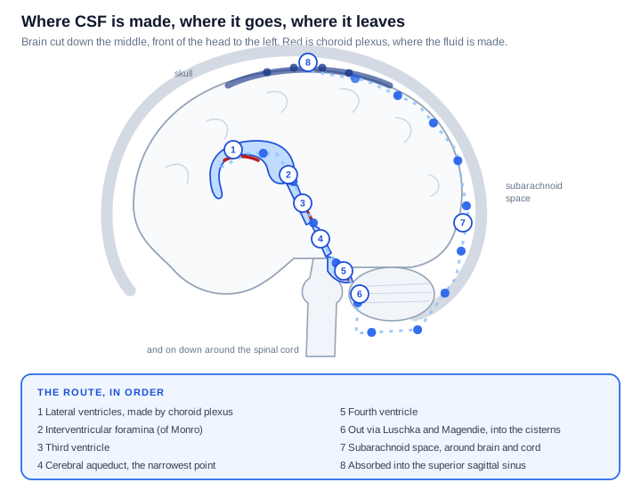
Watching it move makes the shape of the problem clearer than any still picture does.
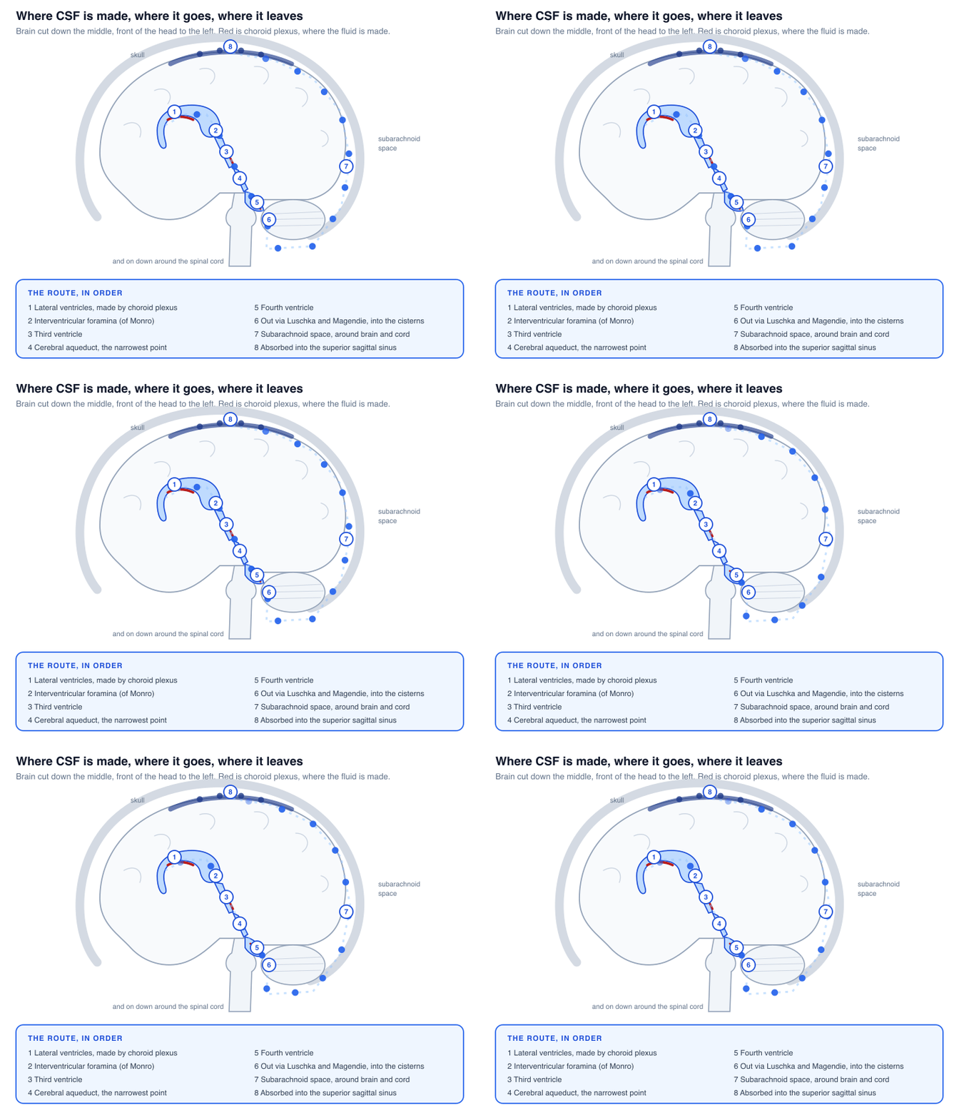
The step worth noticing is the fourth one. The cerebral aqueduct, connecting the third ventricle to the fourth, is the narrowest part of the entire system, a channel a couple of millimetres across carrying everything the choroid plexus produces. Everything upstream of it depends on it staying open.
At the end of the circuit, absorption happens mainly through arachnoid granulations, small tufts that push up into the large venous sinuses draining the skull, letting fluid pass into the bloodstream. Lymphatic drainage, including vessels in the meninges described in 2015, is now understood to carry a share as well, and this part of the story is still being revised.
What it is actually for
It makes the brain weigh almost nothing. This is the big one. By Archimedes' principle the brain displaces its own volume of fluid and is buoyed up by it. Published figures for the resulting effective weight vary quite a bit, commonly between about 25 and 50 grams, because the answer is sensitive to the exact densities used. The principle holds regardless of which figure you take: a 1.4 kg organ ends up weighing a small fraction of that. Without it the brain would deform under its own weight and tear at the structures tethering it.
It absorbs shock. A layer of liquid between brain and skull means a knock decelerates the fluid first.
It gives the brain somewhere to put things. The brain has no conventional lymphatic drainage, so CSF carries away metabolic waste. This clearance is more active during sleep, which is part of why sleep looks increasingly like maintenance rather than downtime.
It buffers pressure. The skull is a rigid box with a fixed volume shared between brain tissue, blood and CSF. If one grows, another has to give, and CSF is the component with the most room to move.
It keeps the chemistry steady. Neurons are sensitive to the ionic environment they sit in, and tightly controlled CSF composition means a meal or a hard workout does not change the fluid the brain is bathing in.
What happens when it goes wrong
Because production does not depend on drainage working, a blockage does not reach a comfortable equilibrium. The fluid keeps arriving.
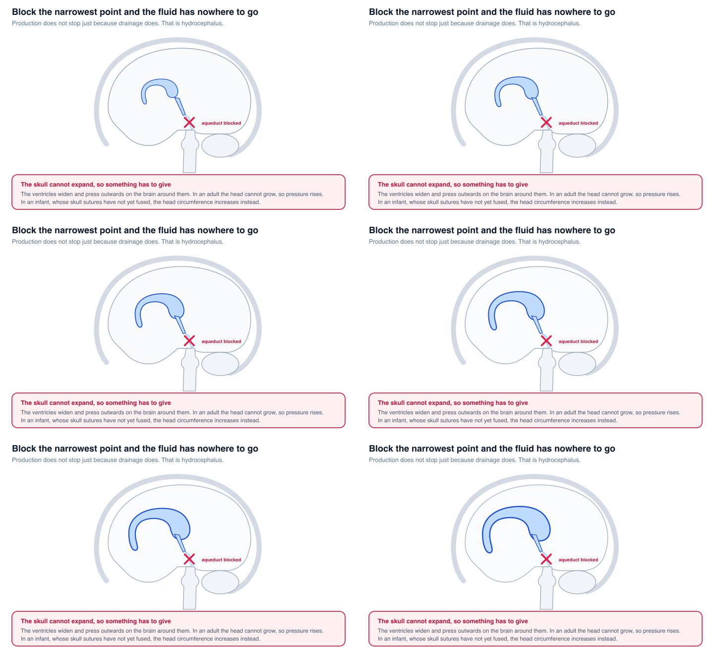
This is hydrocephalus, and it comes in two broad forms. If the obstruction is inside the ventricular system, often at that narrow aqueduct, the fluid cannot get out. If the plumbing is clear but absorption has failed, usually after bleeding or infection has scarred the granulations, the fluid gets out but has nowhere to go.
Age decides what the pressure does next. An infant's skull sutures have not fused, so the head enlarges and the pressure stays lower for longer. An adult skull cannot expand at all, so pressure rises instead, and the classic triad of headache that is worse lying down, vomiting and visual disturbance follows.
There is also a version where the pressure reading comes back normal. Normal pressure hydrocephalus presents with an unsteady gait, urinary incontinence and cognitive decline. It is worth knowing about because it is one of very few causes of dementia-like symptoms that can be treated by draining fluid, and it is often mistaken for something irreversible.
What a lumbar puncture reads
A sample is taken from the lumbar spine for a specific anatomical reason: the fluid-filled sac extends further down the back than the spinal cord does.
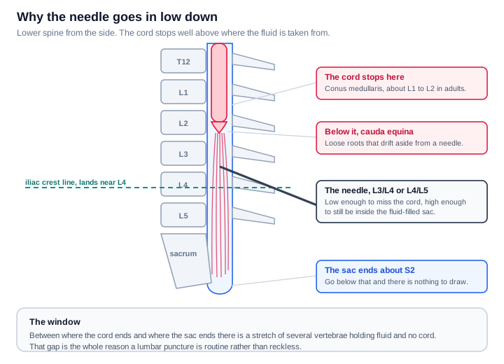
The cord tapers and ends at the conus medullaris, around the L1 to L2 level in adults. The dural sac carries on to roughly S2. Between those two points is a stretch of several vertebrae containing fluid and the loose nerve roots of the cauda equina, which drift aside rather than being skewered. That gap is why the procedure is routine. The needle goes in at L3/L4 or L4/L5, and the line across the top of the hip bones is the landmark used to find the level.
Three things get read, and the first happens before any fluid leaves the room.
The pressure. Measured as it opens, normally somewhere around 10 to 20 cm of water in an adult lying on their side. High suggests infection, bleeding or a mass. Low suggests a leak.
The appearance. Normal CSF is clear and colourless. Cloudy means cells. A yellow tinge, xanthochromia, means blood has been there long enough to break down, which distinguishes a genuine subarachnoid haemorrhage from blood introduced by the needle itself.
The contents. This is where the clean baseline pays off.
| Pattern | Cells | Glucose | Protein | Typically |
|---|---|---|---|---|
| Bacterial meningitis | High, mostly neutrophils | Low | High | A medical emergency |
| Viral meningitis | Moderate, mostly lymphocytes | Normal | Mildly raised | Usually self-limiting |
| Tuberculous or fungal | Lymphocytes | Very low | Very high | Slower onset |
| Subarachnoid haemorrhage | Red cells, xanthochromia | Normal | Raised | Bleed around the brain |
| Guillain-Barré syndrome | Normal | Normal | High | Protein up, cells not |
| Multiple sclerosis | Normal or mild | Normal | Oligoclonal bands | Immune activity inside the CNS |
The glucose column does a lot of work. Bacteria consume glucose and viruses do not, so a low CSF glucose points hard at a bacterial or tuberculous cause rather than a viral one, and it does so before any culture has had time to grow.
The Guillain-Barré row has a name of its own, albuminocytological dissociation, which is a long way of saying the protein went up without the cell count following. Normally those two rise together, so the fact that they have come apart is itself the finding.
More recently the same sample has been used for degenerative disease, where amyloid and tau levels are measured as markers of Alzheimer's pathology.
The short version
Made continuously by the choroid plexus at around half a litre a day, held at about 150 mL, replaced several times daily. Out through the ventricles, down the aqueduct, around the brain and cord, absorbed into the venous sinuses at the top. It makes a 1.4 kg brain weigh almost nothing, cushions it, flushes its waste, and absorbs changes in pressure.
Block it and the pressure has nowhere to go. Sample it and, because it starts out so clean, nearly anything you find in it means something.
Sources and further reading
- Cerebrospinal fluid production rates and turnover, review
- Choroid plexus and CSF formation
- Cotransporter-mediated water transport in CSF formation, Steffensen and colleagues
- CSF normal values and abnormalities, laboratory reference
- Buoyancy and the brain, neutral buoyancy
If you want the wider map of what sits where in the brain, I wrote that up in Which Part of the Brain Does What, and the signalling layer underneath it in The Human Nervous System: A Visual Guide.
Never miss a post
New guides on VMware Cloud Foundation, Aria Suite, and infrastructure automation. Follow the blog in your feed reader and new posts show up as soon as they are published.
Using a different reader? Copy the feed URL and add it there.